Knee Muscle Anatomy Mri / Knee Anatomy Magnetic Resonance Mr Axial. Atlas of knee mri anatomy. An mri of the knee of a healthy subject was performed in the 3 planes of space (coronal, axial, sagittal) commonly used in osteoarticular imaging, with two weightings most commonly used to explore the musculoskeletal pathology of the knee: Knee anatomy is incredibly complex, and problems with any part of the knee anatomy—including the bones, cartilage, muscles, ligaments and tendons—can cause pain. In this presentation mri anatomy biceps femoris muscle. Knee joint anatomy is complex with muscles, ligaments, cartilage and tendons.
This mri knee cross sectional anatomy tool is absolutely free to use. In this presentation mri anatomy biceps femoris muscle. Radiology department of the washington university school of medicine, st. David rubin and robin smithuis. Plantaris acts weakly to plantar flex the foot and flex the knee.
Mri Knee Joint Anatomy from image.slidesharecdn.com In conclusion, we describe the normal mri anatomy of the distal biceps femoris and the relationship of this muscle with the common peroneal nerve. Cross sectional anatomy of the knee based on mri : Plantaris acts weakly to plantar flex the foot and flex the knee. General anatomy and musculoskeletal system. When a muscle has different orientations of the tendons it means that there are different patterns of edema possible depending on the tendon injured. Prescribe sagittal plane off axial images with line parallel to bony glenoid. Anatomy of the knee bones around the knee. Mri knee anatomy knee sagittal anatomy free cross sectional anatomy mri knee mri diagnostic imaging :
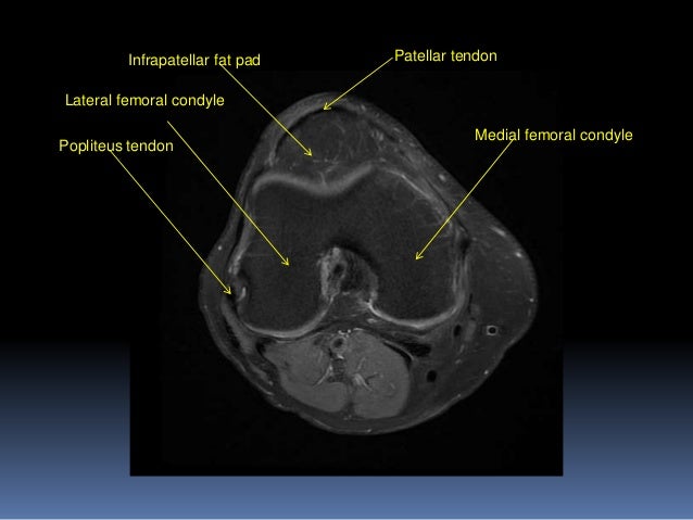
Tibial tuberosity with distal patella tendon insertion.
Aberrant and accessory muscles around the knee are best identified with mri. Plantaris acts weakly to plantar flex the foot and flex the knee. David rubin and robin smithuis. Doctors may recommend a knee mri if a patient experiences the following(3): Atlas of knee mri anatomy. Cross sectional anatomy of the knee based on mri : Cross sectional anatomy of the knee based on mri : A coronal scan goes through the knee, front. 1 november 2002 mri anatomy of the knee and shoulder james y. This mri knee cross sectional anatomy tool is absolutely free to use. Anatomy of the knee bones around the knee. The common peroneal nerve typically courses downward within abundant fat posterior to the short head of the biceps femoris muscle and superficial to the lateral head of the gastrocnemius muscle, but. In this presentation mri anatomy biceps femoris muscle.
Click now to learn more about the bones, muscles, and soft tissues of these regions at leg and knee anatomy: Injuries such as anterior cruciate ligament, meniscus and rotator cuff tears are all easily diagnosed when there is a firm understanding and knowledge of human anatomy. Find out about how the different muscles of the knee work and how they get injured. General anatomy and musculoskeletal system. The images may also help physicians to distinguish normal, healthy tissues from dead tissues(2).
Http Www Smartview Co Wp Content Uploads 2014 02 Imagen Mr Normal Anatomia Rodilla Pdf from Louis, usa and the rijnland hospital in leiderdorp, the netherlands Prescribe sagittal plane off axial images with line parallel to bony glenoid. While a detailed explanation of mri protocols and mr physics is beyond the scope of this text, fast spin echo (fse) mri is most commonly utilized for mri of the knee. Both the pronounced accuracy of the mri and the high prevalence of knee disorders, makes the knee mri the most frequently ordered imaging procedure of the musculoskeletal system. Find out how the different structures fit together in our knee diagram the knee joint is the largest and one of the most complex joints in the human body. Doctors may recommend a knee mri if a patient experiences the following(3): Tips to keep joints healthy. This webpage presents the anatomical structures found on knee mri.
Louis, usa and the rijnland hospital in leiderdorp, the netherlands
The muscles of the knee include the quadriceps, hamstrings, and the muscles of the calf. These motions of the knee allow the body to perform such important movements as walking, running, kicking, and jumping. Naturally, in order to assess pathologic knee imaging, it is necessary to know the appearance of a normal knee mri. Magnetic resonance imaging (mri scan): This section of the website will explain large and minute details of sagittal knee cross sectional. Knee muscle anatomy mri : The muscles of the knee include the quadriceps, hamstrings, and the muscles of the calf. Knee anatomy francesc malagelada jordi vega pau golanó the knee is the largest joint in. 4, infrapatellar fat pad of hoffa. Mri knee joint anatomy 1. Injuries such as anterior cruciate ligament, meniscus and rotator cuff tears are all easily diagnosed when there is a firm understanding and knowledge of human anatomy. The images may also help physicians to distinguish normal, healthy tissues from dead tissues(2). General anatomy and musculoskeletal system.
A coronal scan goes through the knee, front. Tips to keep joints healthy. Both the pronounced accuracy of the mri and the high prevalence of knee disorders, makes the knee mri the most frequently ordered imaging procedure of the musculoskeletal system. Anatomy basic knee mri checklist. The muscles of the knee include the quadriceps, hamstrings, and the muscles of the calf.
Accessory Muscles Of The Knee Radsource from radsource.us There is a flat area of tendon originating from the knee. 1 november 2002 mri anatomy of the knee and shoulder james y. Find out how the different structures fit together in our knee diagram the knee joint is the largest and one of the most complex joints in the human body. Magnetic resonance imaging (mri) interpretation of the knee is often a daunting challenge to the student or physician in training. The main knee muscles are the quadriceps, hamstrings and calf muscles. This mri knee cross sectional anatomy tool is absolutely free to use. Three conventional mri planes that are utilized to evaluate the knee include sagittal (oblique), coronal, and transaxial planes. Song, uc san francisco msiv gillian lieberman md.
1 november 2002 mri anatomy of the knee and shoulder james y.
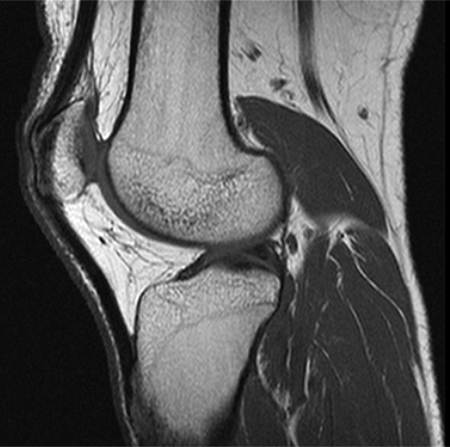
Three conventional mri planes that are utilized to evaluate the knee include sagittal (oblique), coronal, and transaxial planes. Anatomy basic knee mri checklist. 12 photos of the knee muscle anatomy mri. This mri knee cross sectional anatomy tool is absolutely free to use. This webpage presents the anatomical structures found on knee mri. A coronal scan goes through the knee, front. In conclusion, we describe the normal mri anatomy of the distal biceps femoris and the relationship of this muscle with the common peroneal nerve. When a muscle has different orientations of the tendons it means that there are different patterns of edema possible depending on the tendon injured. Mri for evaluating knee pain in older patients: Plantaris can have variable size, but in most cases is difficult to demonstrate on routine mri studies. Knee muscle anatomy mri : Knee joint anatomy is complex with muscles, ligaments, cartilage and tendons. Use the checklist to quiz yourself.
0 Response to "Knee Muscle Anatomy Mri / Knee Anatomy Magnetic Resonance Mr Axial"

0 Response to "Knee Muscle Anatomy Mri / Knee Anatomy Magnetic Resonance Mr Axial"
Post a Comment